You are incorrect - the best interpretation of the chest X ray in our patient is pulmonary edema with a normal heart size.
Your choice: Increased pulmonary vascularity + dilated pulmonary trunk
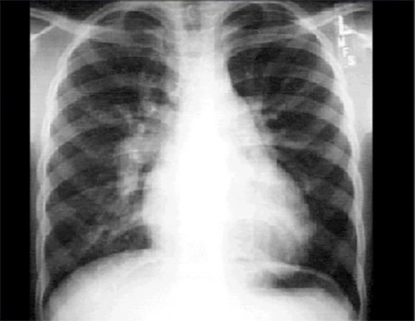
This chest X ray shows increased pulmonary vascularity and a dilated pulmonary trunk.
In this PA view, the pulmonary vasculature markings are clearly visible in the outer third of the lung fields. The convex shadow just beneath the small aortic knob reflects an enlarged main pulmonary artery. Cardiomegaly is also present, as evidenced by the cardiothoracic ratio greater than fifty percent.
These findings are compatible with an intracardiac left-to-right shunt.