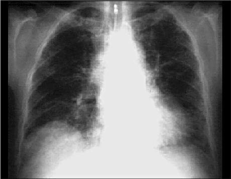
You are incorrect - the best interpretation of the chest X rays in our patient is right ventricular enlargement, prominent pulmonary arteries and decreased peripheral markings.
Your choice: Bilateral pulmonary fibrosis and mild cardiomegaly

This chest X ray shows bilateral pulmonary fibrosis and mild cardiomegaly. This PA view demonstrates
increased diffuse bilateral interstitial pulmonary markings. Note also that the central pulmonary arteries are prominent, compatible with pulmonary hypertension. There is mild cardiomegaly, as evidenced by a slightly increased cardiothoracic ratio. Note the tracheostomy too.