You are incorrect - the best interpretation of the chest X rays in our patient is right ventricular enlargement, prominent pulmonary arteries and decreased peripheral markings.
Your choice: Right ventricular enlargement and left atrial enlargement
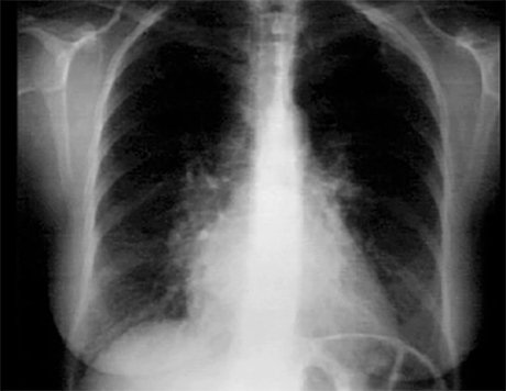
These chest X rays show right ventricular enlargement and left atrial enlargement. The PA view demonstrates left atrial enlargement reflected by the double contour within the heart border, an elevated main stem bronchus and an enlarged left atrial appendage causing straightening of the left heart border. Note also that the cardiothoracic ratio is greater than fifty percent, reflecting cardiomegaly.
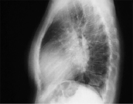
In the lateral view, left atrial enlargement is further reflected by the prominent posterior left atrial shadow. right ventricular enlargement is best seen in this view and is manifested by obliteration of the retrosternal air space.