You are incorrect - the best interpretation of the chest X rays in our patient is right ventricular enlargement, prominent pulmonary arteries and decreased peripheral markings.
Your choice: RV enlargement, dilated pulmonary trunk and ↑ pulmonary vascularity
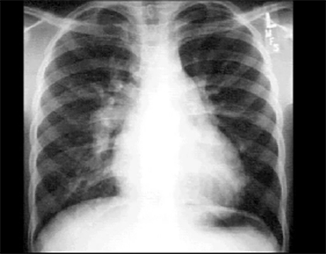
PA
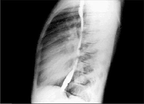
Lat
These chest X rays show right ventricular enlargement, a dilated pulmonary trunk and increased pulmonary vascularity. In this PA view, right ventricular enlargement is suggested by the minimally elevated cardiothoracic ratio and the presence of an upturned apex. The dilated pulmonary trunk
is manifested by the convex density below the aortic knob. There is also prominence of the pulmonary arteries, particularly well seen as dilatation of the right pulmonary artery. Such dilatation is compatible with increased blood flow, as seen with left-to-right shunts.
In the lateral view with barium swallow, right ventricular enlargement is manifested by obliteration of the retrosternal air space. The pulmonary arteries are large.