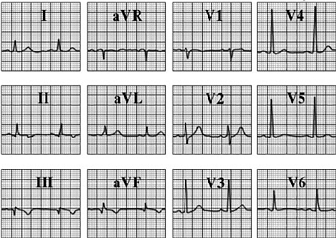
Evolved Inferior Wall Infarction - Post Thrombolysis
This electrocardiogram shows evolution of the inferior wall myocardial infarction. The characteristic features demonstrated here include small Q waves in leads II, III and aVF, with associated symmetric, inverted T waves in these same leads. The ST segments have nearly returned to the baseline. Reperfusion has accelerated these ECG changes in the inferior wall and has resulted in the resolution of the ST-T abnormalities seen in other leads on the initial electrocardiogram. Note also that his type I second degree heart block has resolved and the rhythm is sinus.