You are incorrect - the best interpretation of the electrocardiogram in our patient is right ventricular enlargement + right axis deviation and right atrial enlargement.
Click on the links to learn about this ECG:
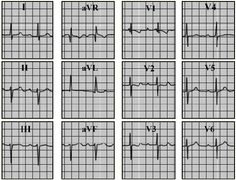
Your choice: Right ventricular enlargement + left axis deviation
This electrocardiogram shows right ventricular enlargement + left axis deviation. The characteristic features of right ventricular enlargement demonstrated here include an rSR' complex and associated ST-T abnormalities in lead V1. Left axis deviation is demonstrated by the predominantly negative QRS complex
in leads II and aVF, with a tall positive complex in lead aVL. First degree AV block is also present, as evidenced by the prolonged PR interval. These electrocardiographic changes are found in ostium primum atrial septal defects.