You are incorrect - the best interpretation of the electrocardiogram in our patient is right ventricular enlargement + right axis deviation and right atrial enlargement.
Click on the links to learn about this ECG:
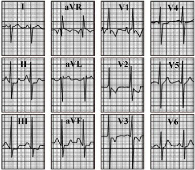
Your choice: Right ventricular hypertrophy + biatrial enlargement
This electrocardiogram shows right ventricular hypertrophy + biatrial enlargement. The characteristic features of right ventricular hypertrophy demonstrated here include a tall R wave in V1 with an R/S ratio of one or more. ST-T wave abnormalities in those leads with tall R waves and right axis deviation reflected in part by the negative QRS complex in lead I. Right atrial enlargement is reflected by the tall P wave in lead II and lends further support to the diagnosis of right ventricular hypertrophy. Left atrial enlargement is reflected by the increased width of the P wave in lead II and the prominent negative component of the P wave in lead V1. The hallmark of right ventricular hypertrophy is the tall R wave
in lead V1. This must be differentiated from the other ECG patterns with similar R waves, such as true posterior wall myocardial infarction.