You are incorrect - our patient's chest X rays are entirely normal.
Your choice: Straight back
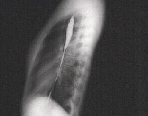
The lateral X ray with barium swallow shown here clearly demonstrates the reduced anteroposterior dimension with a loss of dorsal kyphosis that is, a straight back. And there is obliteration of the
retrosternal space.
Because of this skeletal variation, the cardiac silhouette and pulmonary artery may appear to be enlarged in the PA chest X ray. Patients with this variation may present with an exaggeration of normal bedside findings and mitral valve prolapse may be associated with this syndrome.