You are incorrect - our patient's chest X rays show left atrial enlargement and right ventricular enlargement.
Your choice: Calcified mitral valve annulus
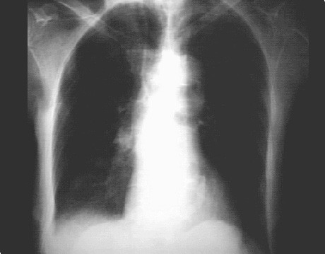
In this overpenetrated PA view, it is demonstrated by the curvilinear density adjacent to the thoracic spine and within the cardiovascular silhouette.
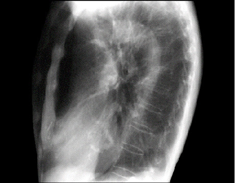
The lateral view is often best for seeing a calcified mitral valve annulus. The “J” shaped density is well seen here, as it does not overlap bony structures.