You are incorrect - our patient's chest X rays show left atrial enlargement and right ventricular enlargement.
Left Ventricular Enlargement + Dilated Aorta PA and Lat
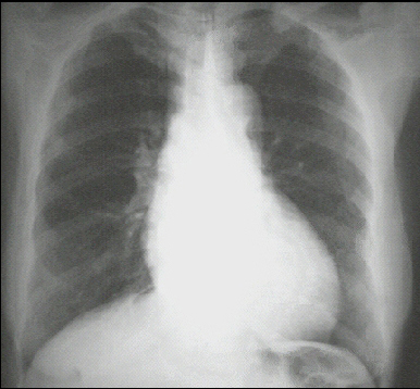
These chest X rays show left ventricular enlargement and a dilated aorta The PA view demonstrates cardiomegaly, as evidenced by a cardiothoracic ratio greater than 50%. Note also the increased inferolateral cardiac border that is consistent with ventricular enlargement due to volume overload. The ascending, transverse, and descending aortic shadows are also prominent.
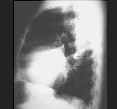
The lateral view shows left ventricular enlargement, as evidenced by posterior displacement of the left ventricular shadow.