You are incorrect - the best interpretation of the electrocardiogram in our patient is right ventricular hypertrophy and biatrial enlargement.
Click on the links to learn about this ECG:
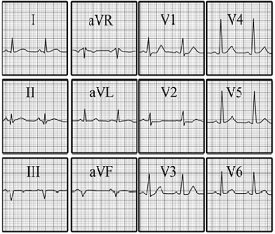
Your choice: Inferoposterior wall myocardial infarction
This electrocardiogram shows an inferoposterior myocardial infarction.
The characteristic features demonstrated here include: Q waves in leads II and aVF, reflecting inferior wall damage; and a tall R wave associated with a positive T wave in lead V1, reflecting posterior wall damage.
Note that the tall R wave in lead V1 may also be seen in patients with right ventricular hypertrophy.