You are incorrect - the best interpretation of the chest X rays in our patient is left ventricular enlargement and a dilated ascending aorta.
Your choice: Calcified left ventricular aneurysm
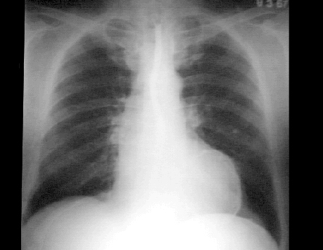
PA
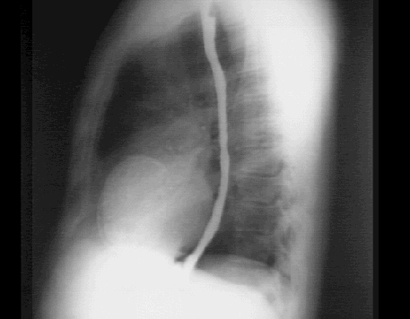
Lateral
These chest X rays show a calcified left ventricular aneurysm.
In this PA chest X ray with barium swallow it is demonstrated at the apex of the left ventricle.
This lateral chest X ray with barium swallow clearly shows the circular density that defines a left ventricular aneurysm.