You are incorrect - the best interpretation of the chest X rays in our patient is cardiomegaly, pulmonary venous congestion and a pleural effusion.
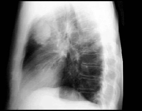
Your choice: Pulmonary Lesion PA and Lat
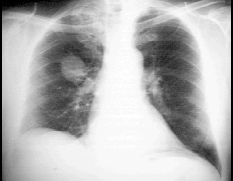
These chest X rays show a pulmonary lesion, or mass. In this PA view, a solitary lesion is seen in the right upper lobe. The mass shows no cavitation or calcification and should be considered cancerous unless proven otherwise. There is also asymmetry of the lung apices with
pleural thickening and stranding on the right. This may be due to radiation or previous granulomatous disease such as tuberculosis. Tha cardiovascular structures are unremarkable. The lateral view also demonstrates the solitary lesion
or mass in the peripheral portion of the anterior segment of the right upper lobe, with pleural thickening and stranding in the apex. The cardiovascular structures are, again, within normal limits.