You are incorrect - our patient's chest X rays show left ventricular enlargement + left atrial enlargement.
Your choice: Left Ventricular Enlargement + Pulmonary Congestion PA and Lat
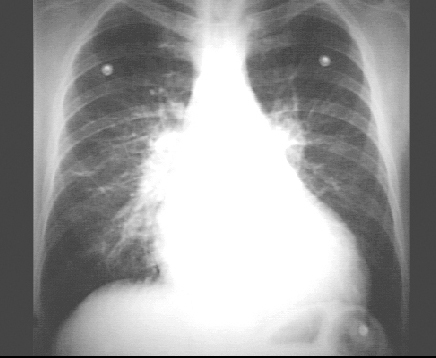
PA view
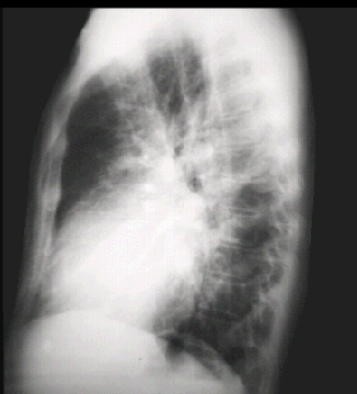
Lateral view
The PA view demonstrates the enlarged left ventricle as an increase in the inferolateral cardiac border, associated with an increased cardiothoracic ratio. Pulmonary congestion is demonstrated by a diffuse increase in vascular markings throughout the lungs. Prominent pulmonary arteries are also seen in the hilar region.
Note the electrode shadows in this patient whose chest X rays were taken while being monitored.
The lateral view shows left ventricular enlargement, as evidenced by posterior displacement of the left ventricular shadow. Note the increased vascular markings in the lung fields.