You are incorrect - the best interpretation of the chest X rays in our patient is pericardial calcification
Your choice: Lung mass and pericardial effusion
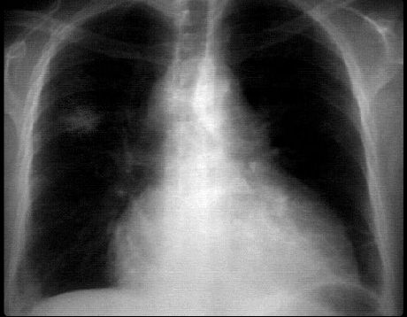
This chest X ray demonstrates a lung mass and pericardial effusion.
The PA view demonstrates a non-calcified mass in the peripheral portion of the right upper lobe.
The cardiopericardial silhouette is enlarged. Whether this is a pericardial effusion and/or cardiac enlargement would require additional imaging studies.
This left hilum is also abnormal from either a mediastinal mass or an enlarged pulmonary artery. With the lung mass, the former is more likely.