You are incorrect - the best interpretation of the chest X ray in our patient is pulmonary venous congestion.
Your choice: Calcified Coronary Arteries Lat and Enlargement
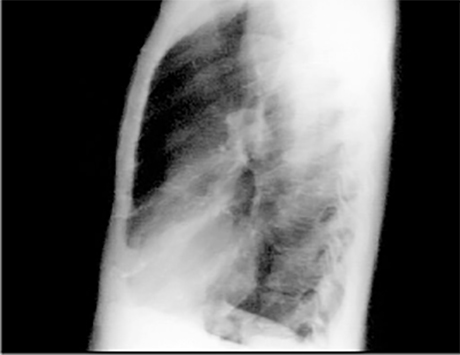
This chest X ray shows calcified coronary arteries.
The lateral view is often best for demonstrating calcium in the coronary vessels. Calcifications are much more common in the proximal parts of the vessel, usually within two centimeters of the root of the aorta and reflect coronary atherosclerosis.
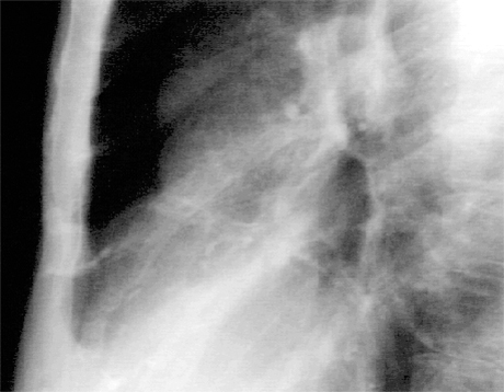
In this enlargement of the lateral view, the railroad track shadows of calcium are more easily seen. There is proximal calcification of some of the branches of the main vessel.
In this enlargement of the lateral view, the railroad track shadows of calcium are more easily seen. There is proximal calcification of some of the branches of the main vessel.