You are incorrect - the best interpretation of the electrocardiogram in our patient is acute anterior wall myocardial infarction.
Click on the links to learn about this ECG:
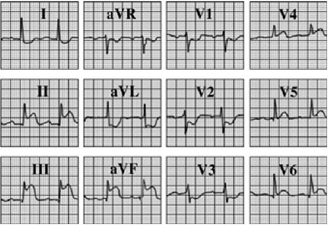
This electrocardiogram shows an acute inferior wall myocardial infarction. The characteristic feature demonstrated here is the marked ST segment elevation in the leads that reflect the inferior wall, that is, leads II, III, and aVF.
Minimal elevation in leads V5 and V6 suggests there is also some lateral involvement.
These ST segment elevations are diagnostic of acute inferolateral transmural injury that almost always evolves to infarction in the absence of early intervention. ST segment depression is present in leads I, aVL, and V1 through V3. This suggests that a significant amount of myocardium is in jeopardy, as patients with these reciprocal changes often have large infarctions.
The PR interval is prolonged, suggesting increased parasympathetic tone and/or possible AV node ischemia, as is commonly seen in inferior wall infarction.